Showing posts with label cell. Show all posts

Wednesday, August 30, 2017

CELL DEATH PROTEINS KEY TO FIGHTINING DISEASE


Melbourne researchers have uncovered key steps involved in programmed cell death, offering new targets for the treatment of diseases including lupus, cancers and neurodegenerative diseases

The research teams from the Walter and Eliza Hall Institute worked together to discover the three-dimensional structure of a key cell death protein called Bak and reveal the first steps in how it causes cell death. Their studies were published in Molecular Celland Proceedings of the National Academy of Sciences.
Programmed cell death, known as apoptosis, occurs naturally when the body has to remove unwanted cells. Chemical signals tell the cell to die by activating the apoptosis proteins Bak and Bax, which break down the 'energy factory' of the cell, known as the mitochondria. When this process goes awry, defective cells such as cancer cells can continue to live, or healthy cells can die unnecessarily, such as occurs in Alzheimer's disease.
Visualizing death proteins
Using the Australian Synchrotron, Mr Jason Brouwer, Dr Peter Czabotar, Dr Ruth Kluck and colleagues from the institute's Structural Biology division investigated how the structure of Bak changes in order to initiate cell death. The research was published in Molecular Cell.
"Understanding the way cell death proteins work and what they look like is crucial to finding new ways to treat disease," Dr Czabotar said. "Our research showed how Bak morphs from one shape to another to trigger apoptosis. Once Bak becomes 'activated' within the cell, it couples with another Bak molecule to form a 'dimer', which then goes on to initiate apoptosis."
Dr Czabotar said understanding apoptosis would allow researchers to develop new ways to treat disease. "Knowing the structure of these proteins and how they work in the cell is essential in designing new treatments to fight disease."
Seeking the hole story
Dr Dana Westphal, Dr Kluck, Dr Grant Dewson, Professor Jerry Adams and colleagues from the Molecular Genetics of Cancer and Cell Signalling and Cell Death divisions examined how the Bak and Bax dimers attach to mitochondria and perforate them. The research was published in Proceedings of the National Academy of Sciences.
Dr Kluck said dimers of Bak and Bax break open the mitochondrial surface, but the mechanism remains poorly understood. "A crucial stage of apoptosis is the release of key proteins from within the mitochondria," she said. "Scientists thought this happened by Bak and Bax poking through the mitochondrial membrane to form a hole, but our work has shown this doesn't happen. Instead, these proteins collapse onto the oily surface of the mitochondria and crowd the surface until holes form."
"We and others are now working to discover exactly how these proteins come together to destroy the mitochondria and trigger apoptosis. A deeper understanding of this pivotal event is likely to suggest new ways to regulate apoptosis to combat disease."


Monday, August 21, 2017

New Drugs To Be Targeted At Pain Receptors Deep In The Nerve Cell


Today's post from sciencedaily.com (see link below) may at first sight seem a little difficult to understand but basically, when the body experiences pain, there are pain receptors on the surface of nerve cells that identify it and transmit the information further and let you feel that pain. If the pain is extreme, sometimes these pain receptors retreat to the nucleus of the cell, as if it's 'safer' there. Pain relieving drugs are designed to block the pain signals in the receptors at the surface of the cell but if the receptors have migrated to the nucleus then the drugs don't have any effect. This knowledge enables researchers to design pain-relieving drugs that can penetrate the nerve cell to the nucleus and thus block the signals from reaching the receptors. They can then 'safely' return to the surface of the cell. At least that's the theory and although it may seem like double-dutch to most of us, sometimes it's interesting to know in which direction the scientists are going.


Location may be key to effectively controlling pain
Date:February 3, 2016 Source:McGill University
 
In real estate, location is key. It now seems the same concept holds true when it comes to stopping pain. New research published in Nature Communications indicates that the location of receptors that transmit pain signals is important in how big or small a pain signal will be -- and therefore how effectively drugs can block those signals.

Blocking pain receptors in the nucleus of spinal nerve cells could more effectively control pain than interfering with the same type of receptors located on cell surfaces. The scientists also found that when spinal nerve cells encounter a painful stimulus, some of the receptors will migrate from the cell surface into the nucleus.

A team of researchers led by McGill University's Director of Anesthesia Research Terence Coderre and Karen O'Malley at Washington University in St. Louis, found that rats treated with investigational drugs to block the activity of the receptors in the nucleus soon began behaving in ways that led them to believe the animals had gotten relief from neuropathic pain. According to Prof. Coderre, "drugs that penetrate the spinal nerve cells to block receptors at the nucleus were effective at relieving pain, while those that don't penetrate the nerve cells were not. Rats with nerve injuries had less spontaneous pain and less pain hypersensitivity after blocking receptors at the nucleus, while the pain sensitivity of normal rats was not affected."

Location is key
Scientists have been studying glutamate receptors in the pain pathway for decades. What's new, Coderre explained, is that these most recent experiments -- in cell cultures and rats -- demonstrate that the location of the receptor in the cell has a major effect on the cell's ability to transmit pain signals.

The researchers focused mainly on nerve cells in the spinal cord, an important area for transmitting pain signals coming from all parts of the body.

"We'll now focus our research at determining what events cause the glutamate receptors to migrate to the nucleus, and how to produce drugs that more specifically block glutamate receptors only at the nucleus," added Coderre.

Story Source:

The above post is reprinted from materials provided by McGill University. The original item was written by Cynthia Lee. Note: Materials may be edited for content and length.

Journal Reference:
Kathleen Vincent, Virginia M. Cornea, Yuh-Jiin I. Jong, André Laferrière, Naresh Kumar, Aiste Mickeviciute, Jollee S. T. Fung, Pouya Bandegi, Alfredo Ribeiro-da-Silva, Karen L. O’Malley, Terence J. Coderre. Intracellular mGluR5 plays a critical role in neuropathic pain. Nature Communications, 2016; 7: 10604 DOI: 10.1038/NCOMMS10604


http://www.sciencedaily.com/releases/2016/02/160203111018.htm

Sunday, August 20, 2017

Cell Toxins That Cause Neuropathy


Today's post from source.wustl.edu (see link below) attempts (successfully) to put some very complex information into words that we as lay readers can pretty much understand. It talks about the toxins produced by Schwann cells which 'leak' out onto adjacent axons and damage them, causing the nerve pain we feel with neuropathy. However, these toxins have been called into action by the mitochondria (energy generators of the cells) which have been damaged themselves and no longer produce the energy needed to make nerve cells work normally. With me so far? Probably not but this article explains the whole process and teaches us how nerves and their complex structures actually work. Finding drugs to block this toxin production is the aim. Have a read. You might surprise yourself how quickly you form a picture of what's going on with neuropathy.
 

New clues to causes of peripheral nerve damage
By Caroline Arbanas March 6, 2013

Anyone whose hand or foot has “fallen asleep” has an idea of the numbness and tingling often experienced by people with peripheral nerve damage. The condition also can cause a range of other symptoms, including unrelenting pain, stinging, burning, itching and sensitivity to touch.

Although peripheral neuropathies afflict some 20 million Americans, their underlying causes are not completely understood. Much research has focused on the breakdown of cellular energy factories in nerve cells as a contributing factor.

Now, new research at Washington University School of Medicine in St. Louis points to a more central role in damage to energy factories in other cells: Schwann cells, which grow alongside neurons and enable nerve signals to travel from the spinal cord to the tips of the fingers and toes.

The finding may lead to new therapeutic strategies to more effectively treat symptoms of this highly variable disorder, the scientists reported March 6 in the journal Neuron.

“We found that a toxic substance builds up in Schwann cells that have disabled energy factories, leading to the same kind of nerve damage seen in patients with neuropathies,” said senior author Jeffrey Milbrandt, MD, PhD, the James S. McDonnell Professor of Genetics and head of the Department of Genetics. “Now, we’re evaluating whether drugs can block the buildup of that toxin, which could lead to a new treatment for the condition.”

The most common cause of peripheral neuropathy is diabetes, which accounts for about half of all cases. The condition also can occur in cancer patients treated with chemotherapy, which can damage nerves.

In the body, Schwann cells wrap tightly around nerve axons, the fibers that relay nerve signals. Graduate student and first author Andreu Viader and colleagues in Milbrandt’s lab studied Schwann cells in mice with genetically disabled mitochondria, or cellular energy factories. Under normal conditions, these mitochondria produce fuel and intermediates of energy metabolism that allow nerve cells to function.

The researchers showed that the crippled mitochondria activated a stress response in the Schwann cells. Instead of synthesizing fatty acids, a key component of Schwann cells, the cells burned fatty acids for fuel.


Over time, inefficient burning of fatty acids by the crippled mitochondria leads to a build up of acylcarnitines, a toxic substance, in the Schwann cells. The researchers found levels of acylcarnitines up to 100-fold higher in these mutant Schwann cells than in healthy Schwann cells.

And the bad news doesn’t end there. Eventually, the toxin leaks out of the Schwann cells and onto the nerve axons. Studying neurons in petri dishes, the researchers showed that acylcarnitines damage nerve axons and disrupt the ability of nerves to relay signals.

“The toxin leaking out of the Schwann cells and onto the adjacent nerve axons causes damage that results in pain, numbness, tingling and other symptoms,” Milbrandt said. “We think that is a likely mechanism to explain the degeneration of axons that is known to occur in peripheral neuropathies.”

The new research suggests that drugs that inhibit the buildup of acylcarnitines may block axonal degeneration. Milbrandt and his team now are evaluating the drugs in mice with disabled Schwann cells to see if they can slow or alleviate the decay of axons. The research is supported by the National Institutes of Health (NIH) Neuroscience Blueprint Center Core Grant (P30 NS057105), the HOPE Center for Neurological Disorders and the National Institutes of Health (AG13730 and PPG 2P01 HL057278).

Viader A, Sasaki Y, Kim S, Strickland A, Workman CS, Yang K, Cross RW, Milbrandt J. Aberrant Schwann cell lipid metabolism linked to mitochondrial deficits leads to axon degeneration and neuropathy. Neuron. March 6, 2013.

Washington University School of Medicine’s 2,100 employed and volunteer faculty physicians also are the medical staff of Barnes-Jewish and St. Louis Children’s hospitals. The School of Medicine is one of the leading medical research, teaching and patient care institutions in the nation, currently ranked sixth in the nation by U.S. News & World Report. Through its affiliations with Barnes-Jewish and St. Louis Children’s hospitals, the School of Medicine is linked to BJC HealthCare.

https://source.wustl.edu/2013/03/new-clues-to-causes-of-peripheral-nerve-damage/

Thursday, August 10, 2017

Stem Cell Lines Released For Neuropathy Research


Today's short but nonetheless important post from healio.com (see link below) announces a breakthrough in future stem cell research for nerve damage. You may have noticed more and more articles appearing over the last two years regarding the successes with stem cell therapy and neuropathy but it's been patchy and generally based on relatively small research studies. Now that a large amount of stem cell lines have been released for research, we can expect much faster progress in this promising line of neuropathy treatment. The article appears to be directed towards lines from the Charcot-Marie-Tooth Association (CMTA) but the widespread ramifications are part of a domino effect in stem cell research. A question of 'watch this space' but progress is being made so hang in there!

New stem cell collection available for neuropathy research
June 22, 2016

 
The New York Stem Cell Foundation Research Institute and the Charcot-Marie-Tooth Association recently announced a new collaboration to make stem cells available to researchers. According to a press release, the stem cell lines represent the largest collection available for a variety of neuropathy disorders of known genetic causation.

The New York Stem Cell Foundation (NYSCF) Research Institute and the Charcot-Marie-Tooth Association (CMTA) originally collaborated in 2014 to collect fibroblasts from patients with Charcot-Marie-Tooth diseases and turn the samples into induced pluripotent stem cell lines. The stem cell lines will be available for investigators through the NYSCF Repository. Of the 19 lines created, 11 are currently available and eight are undergoing reprogramming.

“This important collaboration has resulted in the largest ever number of CMT stem cell lines available for research around the world. This is a milestone for the field,” Susan L. Solomon, chief executive officer and cofounder of NYSCF, said in the release.

Patrick A Livney, chief executive officer of CMTA, said he “is proud of the association’s [Strategy to Accelerate Research] STAR platform, dedicated to finding first treatments for various CMT diseases. As STAR’s progress continues, the CMTA wants to enable and include all stakeholders interested in that mission to join in the effort. This NYSCF collaboration achieves that goal.”

For more information: To order stem cell lines, visit www.nyscf.org/repository.

References:

www.cmtausa.org/

www.nyscf.org/

http://www.healio.com/orthotics-prosthetics/health-care-updates/news/online/%7B7c6b092b-610b-4708-88f9-c53c4fde7d8f%7D/new-stem-cell-collection-available-for-neuropathy-research

Sunday, July 2, 2017

Can a high CD4 cell count cause neuropathic pain


Today's post from EATG: the European Aids Treatment Group, (see link below)is based on a study of 1,539 HIV-patients by the University of California. It reveals much of what most of us already know but interestingly also, the fact that a high CD4 cell count (which we all use as a measure of immune system health) can be a possible cause of neuropathic pain. Not really what we want to hear when we're spending every waking minute trying to boost that very cell count. The article suggests that it may be a consideration when deciding whether to start anti retroviral treatment in the first place. Considering that the current thinking is that it's better all round, to start as soon as possible, the dilemma is obvious. On the other hand, there's not really much of a choice is there? The medication which halts viral development is more or less essential and for some people, the pain may be an unwelcome but necessary by-product.

Debilitating HIV-associated sensory neuropathy remains common
20/05/2010

Debilitating sensory neuropathy remains prevalent in HIV-infected patients, despite a general decline of neurological complications with use of combination antiretroviral therapy.

This finding is from a study in which the researchers tested 1,539 HIV-infected individuals for clinical signs of neuropathy and neuropathic pain.

"We were surprised by the high prevalence," lead author Dr. Ronald Ellis of the University of California, San Diego, told Reuters Health by e-mail.

"Painful neuropathy frequently persists and requires ongoing management," even when antiretroviral therapy has reduced viral load and restored immune function, he said.

In the May Archives of Neurology, Dr. Ellis and colleagues report that 881 patients (57.2%) had HIV-associated sensory neuropathy, and 335 of those 881 (38.0%) had neuropathic pain.

"Neuropathic pain was significantly associated with disability in daily activities, unemployment, and reduced quality of life," the investigators say.

Patients currently taking combination antiretroviral therapy had an adjusted odds ratio of 1.60 for clinical signs of sensory neuropathy, compared to past users or never users of antiretroviral combinations.

Age (aOR 2.13), past use of stavudine, didanosine, or zalcitabine (aOR 1.95), and lower CD4 nadir (aOR 1.16) were also associated with sensory neuropathy.

Paradoxically, neuropathic pain was associated with a higher CD4 nadir, as well as with past use of stavudine, didanosine, or zalcitabine.

In an editorial, Drs. Dennis Kolson and Francisco Gonzalez-Scarano, both of the University of Pennsylvania in Philadelphia, said the "association between neuropathic pain and a higher CD4 nadir suggests that a functional immune system may contribute to the induction of pain." They also suggest considering the risk of neuropathy - and the disability that results -- when deciding whether to start antiretroviral therapy.

Regarding potential for peripheral nerve regeneration and recovery, Dr. Ellis told Reuters Health, "It may be that treatments can be designed to enhance regeneration. Alternatively, we may find that earlier HAART therapy using agents without neurotoxicity can protect individuals from developing neuropathy in the first place."

By Dave Levitan
Medscape Today

http://eatg.org/eatg/Global-HIV-News/Treatment/Debilitating-HIV-associated-sensory-neuropathy-remains-common

Saturday, July 1, 2017

Here Stem cell treatment for sciatic nerve damage


In this posting nearly everybody would help you to get a useful reference based upon study associated with present content articles Stem cell treatment for sciatic nerve damage prospect of debate for the reason that enough many people that happens to be seeking out which will. during personal reference Getting people take advantage of a variety of serps take a look at pics which can be strongly related Stem cell treatment for sciatic nerve damage .

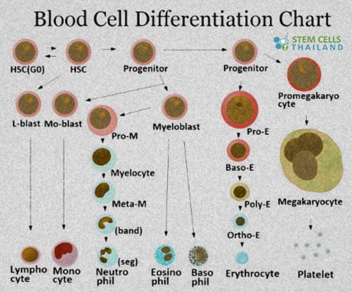
Regenerative Healthcare Clinic for Mesenchymal Stem Cell Therapies Contact Stem Cells Center of Thailand Treat Spinal Cord Injuries With Stem Cells Directed Cell Differentiation Process and Examples meniscus-tear-treatment-arthritis-prp-treatment-in-bangkok-overview Granulocyte Colony-Stimulating Factor G-CSF Stimulation

Pictured preceding you could download and install together with save you this to the pc hard disk making sure that at any time you need it is exclusively levied quite simply. Stem cell treatment for sciatic nerve damage certainly is the primary for your needs exactly who loves to linked issues. Thus most of us had taken this project to accumulate the results with regard to the advantage of the site visitors. Book mark each of our site that will will allow you to search for a great deal more articles or reviews related to the keywords

Stem cell treatment for sciatic nerve damage - for helping establish the interest our targeted visitors are likewise satisfied to earn this page. bettering the grade of this article is going to most people test in the future so you can in fact appreciate after reading this post. In conclusion, this isn't a small number of sayings that really must be which is designed to force one. although a result of the disadvantages connected with dialect, we are able to just existing the actual Stem cell treatment for sciatic nerve damage topic together these


Sunday, June 4, 2017

The Make Up Of A Nerve Cell


Today's very helpful post from cerebromente.org (see link below) gives you everything you ever wanted to know about the make up of a nerve cell. It's fascinating reading although much of it may leave you feeling a sense of information-overload. The point is that everywhere we go in search of information about neuropathy, we will come across several of the terms shown here below. Understanding precisely what they mean, what they do and where they are in relation to each other, increases our knowledge as to what's happening within our nervous system. Doctors don't have the time to explain it all to you but will surely appreciate it if you bring your knowledge to the conversation!


Parts of the Nerve Cell and Their Functions
Silvia Helena Cardoso, PhD


[1. Cell body] [2.Neuronal membrane] [3.Dendrites] [4. Axon] [5. Nerve ending]


1. Cell body

The cell body (soma) is the factory of the neuron. It produces all the proteins for the dendrites, axons and synaptic terminals and contains specialized organelles such as the mitochondria, Golgi apparatus, endoplasmic reticulum, secretory granules, ribosomes and polysomes to provide energy and make the parts, as well as a production line to assemble the parts into completed products. Cytosol - Is the watery and salty fluid with a potassium-rich solution inside the cell containing enzymes responsible for the metabolism of the cell.

1. Nucleus - Derived from the Latin word for "nux", nut, the nucleus is the archivist and the architect of the cell. As archivist it contains the genes, consisting of DNA which contains the cell history, the basic information to manufacture all the proteins characteristic of that cell. As architect, it synthesizes RNA from DNA and ships it through its pores to the cytoplasm for use in protein synthesis.

The.Nucleolus is an organelle within the nucleus which is involved actively in ribosome synthesis and in the transfer of RNA to the cytosol.

2. Golgi Apparatus - membrane-bound structure that plays a role in packaging peptides and proteins (including neurotransmitters) into vesicles.


3. Polyribosomes - there are several free ribosomes attached by a thread. The thread is a single strand of mRNA (messenger RNA, a molecule involved in the synthesis of proteins outside the nucleus). The associated ribosomes work on it to make multiple copies of the same protein.

4. Neuronal membrane (see next box)

5. Mitochondrium - this is the part of the cell responsible for the supply of energy in the form of ATP (adenosine triphosphate). Neurons need an enormous amount of energy. The brain is one of the most metabolically active tissues in the body. In man, for example, the brain uses 40 ml of oxygen per minute. Mitochondria use oxygen and glucose to produce most of the cell's energy.
The brain consumes large amounts of ATP. The chemical energy stored in ATP is used to fuel most of the biochemical reactions of the neuron. For example, special proteins in the neuronal membrane use the energy released by the breakdown of ATP into ADP to pump certain substances across the membrane to establish concentration differences between the inside of the neuron and the outside.

6. Rough Endoplasmic Reticulum and Smooth Endoplasmic Reticulum (7) - A system of tubes for the transportation of materials within the cytoplasm. It may have ribosomes (rough ER) or no ribosomes (smooth ER). With ribosomes, the ER is important for protein synthesis.

Nissl Bodies - Groups of ribosomes used for protein synthesis.


2. Neuronal Membrane


The neuronal membrane serves as a barrier to enclose the cytoplasm inside the neuron, and to exclude certain substances that float in the fluid that bathes the neuron. The membrane with its mosaic of proteins is responsible for many important functions:


- keeping certain ions and small molecules out of the cell and letting others in,
- accumulating nutrients, and rejecting harmful substances,
- catalyzing enzymatic reactions,
- establishing an electrical potential inside the cell,
- conducting an impulse
- being sensitive to particular neurotransmitters and modulators .

The membrane is made of lipids and proteins - fats and chains of aminoacids. The basic structure of this membrane is a bilayer or sandwich of phospholipids, organized in such a way that the polar (charged) regions face outward and the non polar regions face inward.

The external face of the membrane contains the receptors, small specialized molecular regions which provide a kind of "attachment port" for other external molecules, in a scheme analogous to a a key and a keyhole. For each external molecule there is a corresponding receptor. Whenever receptors become attached to a molecule, some alterations of the membrane and in the interior of the cell ensue, such as the modification of permeability to some ions.

3. Dendrites


These structures branch out in treelike fashion and serve as the main apparatus for receiving signals from other nerve cells. They function as an "antennae" of the neuron and are covered by thousands of synapses. The dendritic membrane under the synapse (the post-synaptic membrane) has many specialized protein molecules called receptors that detect the neurotransmitters in the synaptic cleft. A nerve cell can have many dendrites which branch many times, their surface is irregular and covered in dendritic spines which are where the synaptic input connections are made.

4.
Axon 


Axon


Usually a long process which often projects to distant regions of the nervous system. The axon is the main conducting unit of the neuron, capable of conveying electrical signals along distances that range from as short as 0.1 mm to as long as 2 m. Many axon split into several branches, thereby conveying information to different targets. Many neurons do not have axons. In these so-called amacrine neurons, all the neuronal processess are dendrites. Neurons with very short axons are also found. The axons of many neurons are wrapped in a myelin sheat, which is composed of the membranes of intersticial cells and is wrapped around the axons to form several concentric layers. The myelin sheath is broken at various points by the nodes of Ranvier, so that in cross section it looks like a string of sausages. The myelin protects the axon, and prevents interference between axons as they pass along in bundles, sometimes thousands at time.

The cells that wrap around peripheral nerve fibers - that is, nerve fibers outside of the brain and spinal cord - are called Schwann cells (because they were first described by Theodor Schwann). The cells that wrap around axons within the central nervous system (brain and spinal cord) are called oligodendrocytes. The axon, with its surrounded sheath, is called a nerve fiber. Between each pair of sucessive Schwann cells is a gap of a node of Ranvier.


The Axon Hillock

The axon hillock is where the axon is joined to the cell. It is from here that the electrical firing known as an action potential usually occurs.

5. Nerve Ending (Presynaptic Terminals)


Synapses are the junctions formed with other nerve cells where the presynaptic terminal of one cell comes into 'contact' with the postsynaptic membrane of another. It is at these junctions that neurons are excited, inhibited, or modulated. There are two types of synapse, electrical and chemical. Electrical synapses occur where the presynaptic terminal is in electrical continuity with the postsynaptic. Ions and small molecules passing through, thus connecting channels from one cell to the next, so that electrical changes in one cell are transmitted almost instantaneously to the next. Ions can generally flow both ways at these junctions i.e. they tend to be bi-directional, although there are electrical junctions where the ions can only flow one way, these are know as rectifying junctions. Rectifying junctions are used to synchronise the firing of nerve cells.
Chemical synaptic junction is more complicated. The gap between the post- and presynaptic terminals is larger, and the mode of transmission is not electrical, but carried by neurotransmitters, neuroactive substances released at the presynaptic side of the junction. There are two types of chemical junctions. Type I is an excitatory synapse, generally found on dendrites, type II is an inhibitory synapse, generally found on cell bodies. Different substances are released at these two types of synapse. The direction of flow of information is usually one way at these junctions.
Each terminal button is connected to other neurons across a small gap called a synapse. The physical and neurochemical characteristics of each synapse determines the strength and polarity of the new input signal. This is where the brain is the most flexible, and the most vulnerable. Changing the constitution of various neurotransmitter chemicals can increase or decrease the amount of stimulation that the firing axon imparts on the neighbouring dendrite. Altering the neurotransmitters can also change whether the stimulation is excitatory or inhibitory.
http://www.cerebromente.org.br/n07/fundamentos/neuron/parts_i.htm


NEW MECHANISM AFFECTING CELL MIGRATION FOUND


Cell migration is important for development and physiology of multicellular organisms. During embryonic development individual cells and cell clusters can move over relatively long distances, and cell migration is also essential for wound healing and many immunological processes in adult animals. On the other hand, uncontrolled migration of malignant cells results in cancer invasion of metastasis.
Cell migration has mainly been studied in cell culture environment. However, in animal tissues cells predominantly migrate in a three-dimensional environment, where they have to push through adjacent cell-layers and extracellular matrix. Migrating cells are known to form dynamic protrusions at their leading edge, but the function of these actin-rich protrusions has remained elusive.
By using fruit fly as a model system, Minna Poukkula working at the Institute of Biotechnology, University of Helsinki, has found out how actin-rich protrusions contribute to cell migration in animal tissues. She revealed that GMF, a protein that promotes the disassembly of branched actin filament networks, controls the size and lifetime of protrusions in border cell clusters migrating in fruit fly egg chambers. Importantly, diminished protrusion dynamics in GMF-deficient flies correlated with problems in border cell cluster migration.
"These findings demonstrate that efficient actin filament disassembly by GMF is essential for rapid dynamics of cell protrusions, and that this dynamics are important for cell migration in a three-dimensional tissue environment," says Minna Poukkula from the research group of professor Pekka Lappalainen.


Friday, June 2, 2017

The Rapid Rise Of Stem Cell Research For Nerve Pain


Today's post from consultqd.clevelandclinic.org (see link below) takes a rather complex look at developments in stem cell research for neuropathic pain and concludes that significant progress is being made. It's still at the rodent-lab-testing stage but seems much closer to a successful outcome than many studies that we read about. The article is not particularly easy reading for the layman but you can get the gist of what's being discussed. Yet again, this is evidence of the spin-off potential of the current 'war on opioids', which seems to have shaken up the pharmaceutical industry research departments, to the point where they are seriously investigating new possibilities in anticipation of draconian laws restricting opioid distribution. Interesting article.


Stem Cell Therapy for Neuropathic Pain: New Findings Show Promise
Animal studies demonstrate effectiveness
 
Consult QD Aug. 10, 2016

Stem cell research at Cleveland Clinic could pave the way for an entirely new approach to chronic pain treatment that reduces medicine’s current reliance on opioid therapy for intractable pain. The modality also shows promise as a tool to reverse opioid tolerance (OT) and opioid-induced hyperalgesia (OIH), particularly problematic side effects of opioid therapy. Jianguo Cheng, MD, PhD, and his colleagues at Cleveland Clinic have developed patented methods of attenuating opioid tolerance.

Animal studies by Dr. Cheng and his colleagues have demonstrated the effectiveness of mesenchymal stem cell (MSC) transplantation in reducing hyperalgesia due to nerve injury. The group’s work has shown MSC transplantation’s effectiveness in reducing pain induced by sciatic nerve injury in rats and mice. MSC transplantation significantly reduced pain sensitivity evaluated by foot withdrawal thresholds in animals in response to thermal or mechanical stimulation. These cells produced immune modulatory and anti-inflammatory effects, promoted sensory nerve repair, and showed strong analgesic properties that could provide a safer and more effective alternative to current treatment modalities, in the management of neuropathic pain, says Dr. Cheng, Professor of Anesthesiology and Director of the Cleveland Clinic Multidisciplinary Pain Medicine Fellowship Program.

Pain medicine researchers are searching for an alternative to opioid therapy because neuropathic pain often does not respond to morphine and other opioids. Opioid analgesics can also lead to a variety of complications, ranging from itching and constipation to dependence, addiction, respiratory depression and death.

About 30 percent of neuropathy cases are caused by nerve damage associated with diabetes. However, hundreds of diseases are linked to neuropathic pain. Sources of neuropathic pain include alcoholism, amputation (which can result in phantom pain), some chemotherapy drugs (for example, Cisplatin®, Paclitaxel®, Vincristine®), radiation therapy, complex regional pain syndrome type II, trigeminal neuralgia, shingles, spinal stenosis, and central nervous system disorders, such as Parkinson disease and multiple sclerosis.

Recent research by Dr. Cheng and his group has yielded new discoveries that bode well for MSC transplantation as a potential future treatment modality. One investigation compared the analgesic effects of MSC derived from bone marrow with MSC derived from adipose tissue. Adipose-derived cells were found to be as efficacious as bone marrow-derived cells in reducing neuropathic pain in rats. The finding suggests that stem cell therapy could offer a practical option because stem cells from adipose tissue are relatively easy to obtain.

Recent investigations by Dr. Cheng and his colleagues comparing the analgesic effectiveness of intrathecal versus intravenous methods of MSC transplantation show both methods to be equally effective. The finding has important implications because intravenous transplantation of MSC could offer a safer and more expeditious route of delivery than intrathecal transplantation.

“We originally thought that stem cells would have to be introduced intrathecally in order to reduce pain, and that stem cells introduced intravenously would pass through the lungs and fail to produce analgesia,” says Dr. Cheng. “The finding that intravenous transplantation is as effective as intrathecal transplantation is encouraging.”

Dr. Cheng’s group has also discovered that MSCs can be found in the area surrounding the injured nerve following MSC transplantation. “For reasons we do not yet fully understand, these cells have the ability to migrate to the injury site to promote repair of the injured nerve fibers,” Dr. Cheng says. “The cells can sense the injury’s location and travel to it.”

Although many questions must be answered before it can be known whether stem cell therapy is safe and effective for humans, some small patient studies show potential, Dr. Cheng says. According to one observational study in Australia, MSC transplantation reduced pain in patients suffering from trigeminal neuralgia, a particularly difficult condition to treat. “Though the findings are preliminary, the study provides some evidence that what we have learned in the laboratory can be translated to clinical use,” Dr. Cheng says.

Dr. Cheng’s team has achieved analgesia with MSC transplantation from rats to mice, providing early evidence that stem cells’ anti-inflammatory and immuno-modulatory properties can be transferred between species. An important pre-clinical study will be to see whether the transplantation of human stem cells to animals also can produce analgesic and anti-tolerance effects, Dr. Cheng says.

Dr. Cheng’s team presented research at the 2016 annual meeting of the American Academy of Pain Medicine showing MSC’s potential to reverse opioid tolerance and opioid-induced hyperalgesia, problems that can compromise the safety and efficacy of opioid therapy. Intravenous transplantation of bone marrow-derived MSC significantly attenuated OT and OIH in animals whether the transplantation was performed seven days before or 14 days after the initiation of daily morphine injections. These data demonstrate that MSC transplantation can not only prevent the development of OT and OIH but can also reverse it.

https://consultqd.clevelandclinic.org/2016/08/stem-cell-therapy-neuropathic-pain-new-findings-show-promise/?utm_campaign=crowdfire&utm_content=crowdfire&utm_medium=social&utm_source=twitter